WHAT IS HEMANGIOSARCOMA?
Hemangiosarcoma is one of the deadliest forms of canine cancer. Hemangiosarcomas are tumors that are thought to arise from the bone marrow and areas with a rich blood supply. Because tumors tend to grow in these blood-rich areas such as the spleen and heart, they can suddenly rupture, causing massive blood loss, and forcing owners and veterinarians to make difficult decisions within minutes of diagnosis.


WHO DOES HEMANGIOSARCOMA MOST COMMONLY AFFECT?
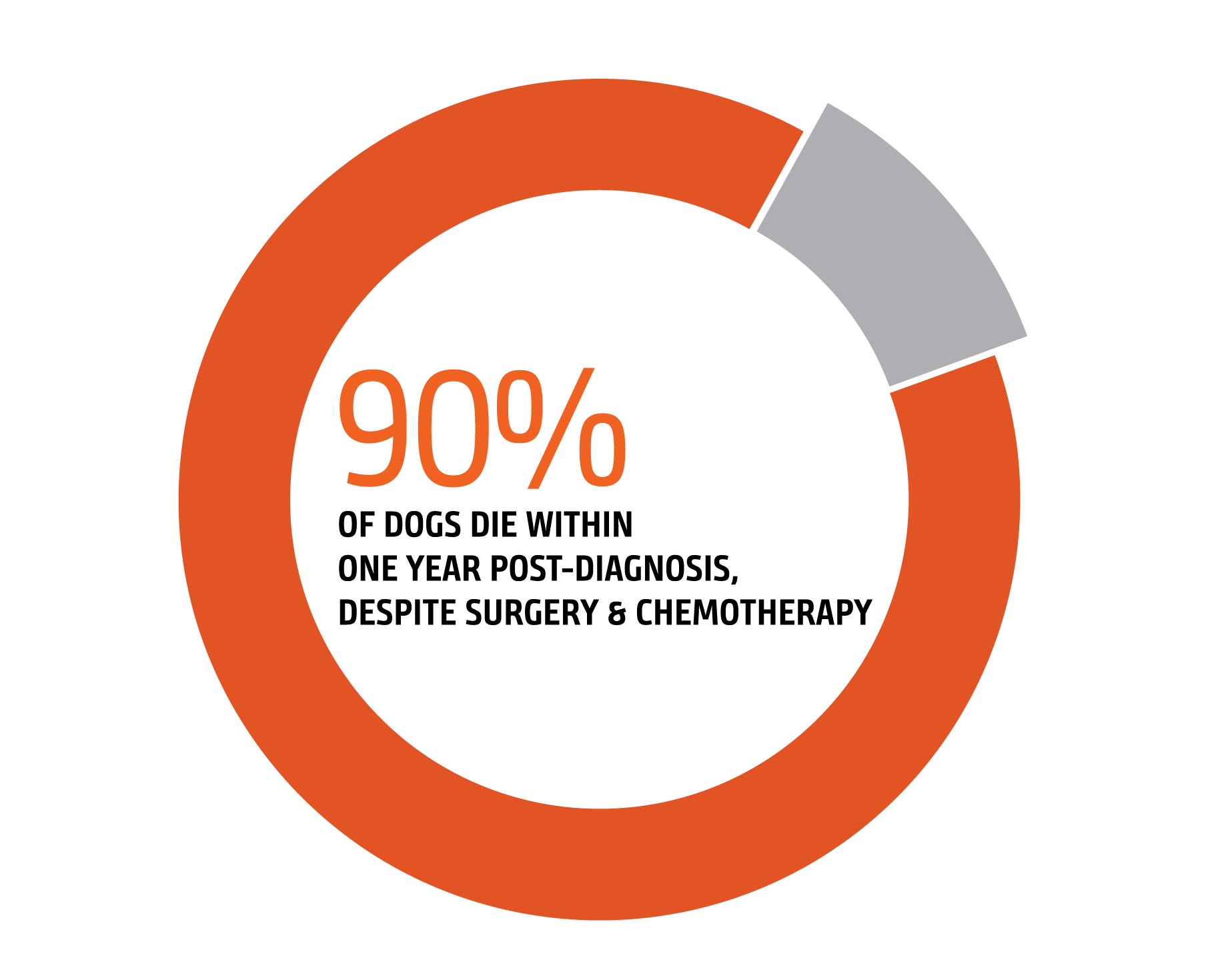
Middle-aged to older dogs; German shepherds, golden retrievers, Portuguese water dogs and Labrador retrievers appear to be at higher risk although any dog can develop hemangiosarcoma. Even when a tumor is quickly detected and removed, the outlook for dogs with hemangiosarcoma is grim.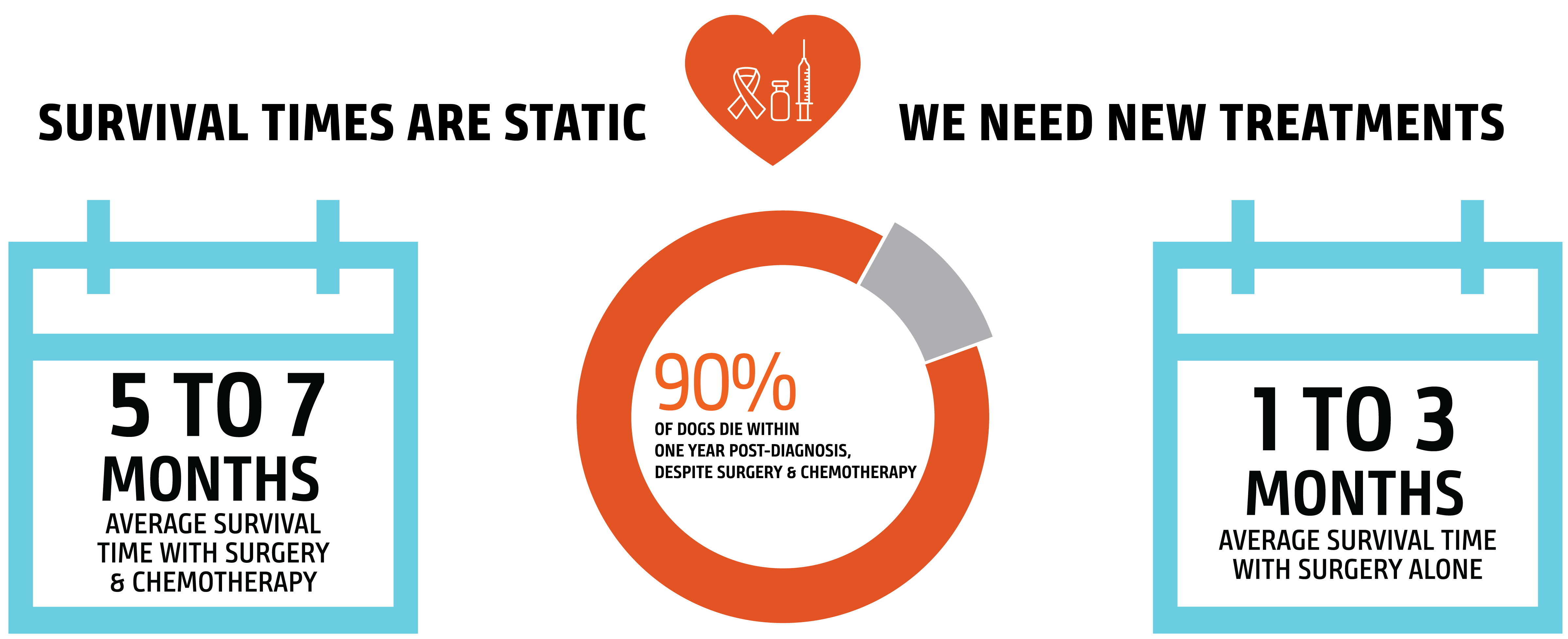


WHAT ARE THE CLINICAL SIGNS?
Unfortunately, no clinical signs (symptoms) are classic for hemangiosarcoma other than sudden, profound, internal bleeding. Other clinical signs reported by owners include: intermittent lethargy or fatigue, anorexia, panting, sudden collapse, sudden death.WHAT DIAGNOSTICS ARE PERFORMED?
Hemangiosarcoma is most often diagnosed by examining tissue collected during surgery or by biopsy, regardless of location within the body. Other tools that are useful in providing a definitive diagnosis and also assessing extent of disease include X-Rays, CT scans and surgery. Often, an echocardiogram is recommended to determine if there is a mass in the heart. |
OUR PROGRESS SO FARMorris Animal Foundation has invested more than $3 million in 20+ years of research to improve the quality and duration of life for dogs diagnosed with hemangiosarcoma. Our funded research has focused on: |
DISEASE BASICSUnderstanding the basic biology of hemangiosarcoma may open the door to new diagnostics, treatments and prevention. |
CHEMOTHERAPY RESISTANCEUnderstanding why hemangiosarcoma becomes drug resistant could improve treatment success as well as identify new chemotherapeutic agents. |
GENETIC LINKSStudying breeds commonly affected by hemangiosarcoma could lead to new diagnostic testing and clues to the role genetics plays in the development of this disease. |
 |
| At Morris Animal Foundation, we want to change the odds for dogs diagnosed with hemangiosarcoma. But we need your help! Our new Hemangiosarcoma Initiative dedicates critically needed funding, people and resources toward new and innovative approaches to diagnose, treat, and possibly cure, this devastating cancer. |
 |
| You can make a difference with your support of this life-changing project. | ||
|
Resources For Grieving Pet Parents
Have you lost a loved one to cancer? Losing a beloved pet is difficult, but knowing there are resources available, and realizing you’re not alone when it comes to pet loss and grief, can make the process just a little easier. Find some resources below to help guide you on this journey.


YOU'RE NOT ALONE: HEMANGIOSARCOMA STORIES

Beating the Cancer Odds
Chase Manhattan beat the odds and survived a hemangiosarcoma diagnosis. Read about her amazing story of survival.
In Heart of Darkness of the Canine Cancer World, Research is a Shining Ray of Hope
Carol Everett tells the story of her dog Davy, a therapy dog who's mission was to bring love to those in need, and his struggle with hemangiosarcoma.

A Dog That Was Meant to Be
Learn how Emily Lucibello honors the memory of her dog Mea through our Loyal Friend program as well as participating in the Golden Retriever Lifetime Study.

Caring For Study Members is This Participant’s Mission
Study participant Gail Ingrish talks about her experience as Study participant and volunteer.