Updated February 1, 2024 – Recognizing American Heart Month, celebrated every February, is a great way to think about all the love our pets give us and offer them. That love fills our hearts, which are both extraordinary and complex organs.
This series delves into heart function, heart diseases affecting dogs and cats and essential tips for owners to identify heart disease in their pets.
A Dynamic, Complicated Organ
The heart, a specialized muscle, fills with blood and contracts to pump it throughout the body. It comprises valves, which help direct blood flow in the proper direction, an electrical system that provides the signals to direct the heart muscle to contract in a regular and orderly fashion and thousands of blood vessels, from the large and powerful aorta to the tiniest capillaries, serve as the conduits for blood as it travels throughout the body.
Heart Parts: Valves, Wall, Nerves and Chambers
Before understanding function, we need a crash course in the different parts of the heart. They are:
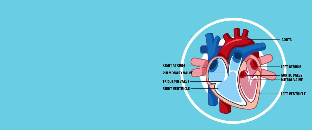
- Valves: The heart has four valves: pulmonic, tricuspid, mitral and aortic.
- Wall: The wall of the heart has three layers: epicardium, myocardium (specialized muscle) and endocardium.
- Nerves: The heart has a defined system of nerve conduction that helps coordinate heart function.
- Chambers: The heart has four chambers: right atrium, right ventricle, left atrium and left ventricle.
Turn the Beat Around – Heart Function Simplified
The heart’s primary role is to pump blood, and the process starts with nerve impulses that trigger the heart muscle to contract. These nerve impulses arise from the autonomic nervous system, and as the name implies, this system is responsible for many essential body functions that happen automatically or without conscious thought.
A nerve impulse travels through the heart, beginning a coordinated muscle contraction. Any disturbance in the impulse transmission can result in an irregular heartbeat – an arrhythmia, which can alter the heart’s ability to function.
The electrical impulse starts in the heart's upper right chamber (the right atrium) at the sino-atrial node, which acts like the heart’s pacemaker. The signal then travels via the conduction system to other areas of the heart. The result is a coordinated series of muscle contractions that result in the expulsion of blood from the heart.
Inside the heart, four valves help blood flow in the proper direction. This simple diagram excellently visualizes the heart’s chambers and blood flow (depicting the right side on the left and the left side on the right by convention).

Blood flows through the heart in a defined pathway:
- Blood enters the right atrium from the body. This blood has low levels of oxygen and high levels of carbon dioxide.
- Blood flows through the tricuspid valve into the right ventricle and then leaves the right ventricle to go to the lungs through the pulmonic valve.
- Carbon dioxide is released into the lungs when we exhale, and oxygen enters the blood when we inhale.
- Blood returns to the heart and enters the left atrium.
- Blood then flows through the mitral valve into the left ventricle.
- Blood then leaves the heart through the aortic valve and is distributed to the rest of the body.
A Quick Overview of Heart Diseases in Dogs and Cats
Various factors can comprise heart function over time. We categorize diseases by their impact on muscles, valves, blood vessels and electrical conduction.
Cardiomyopathy
This general term covers diseases affecting the heart muscle, which can take several forms. For example, dilated cardiomyopathy is a type of heart disease characterized by a thinning of the heart muscle and a decrease in the heart’s ability to contract. This type is most common in large-breed dogs, especially in certain breeds such as Doberman pinschers, Great Danes and Irish wolfhounds, although many breeds are known to develop the disease. DCM tends to affect middle-aged to older dogs and is treatable if caught early. Less commonly, DCM can be the result of certain chemotherapy drugs and nutritional abnormalities.
In contrast, hypertrophic cardiomyopathy thickens the heart walls. Although initially, the heart can contract normally, as the muscle thickens, the heart is less able to contract. Less blood enters the heart, so output decreases. Eventually, the heart muscle fails. There are many intermediaries and combinations of heart muscle disease, but all forms begin as abnormalities of the muscular structures of the heart.
In cats, hypertrophic cardiomyopathy is not only the most common type of heart muscle problem diagnosed but also the most common heart disease of cats overall. HCM also thickens the heart muscle. Cats with HCM are also prone to forming abnormal blood clots, which block major blood vessels, cutting blood supply.
There is a genetic component implicated in some cat breeds (for example, Maine coon cats), but many mixed-breed cats also suffer from this problem. As with all types of heart disease, early diagnosis is the key to successful treatment and longer, healthier lives.
Arterial Thromboembolism
The formation of large, abnormal blood clots, known as arterial thromboembolism, is a common problem in cats with cardiomyopathy. The most common place for a large clot to lodge is in the distal aorta, as it splits into the main vessels that supply each leg, often called a saddle thrombus. This results in a loss of blood flow to both rear legs, resulting in extreme pain and loss of movement in the hind legs.
Other organs, such as the kidneys and brain, can also be affected by abnormal blood clots. Although treatable, prevention of clot formation through specific treatment of the clot and treatment of the underlying heart problem is critical.
Valves and Abnormal Blood Flow
The classic “lub-dub” sound we associate with a heartbeat is the sound of valves closing. Turbulence causes a murmur. Although we think of valve problems as a common cause of murmurs – and they are – it’s also important to remember that murmurs are not always a sign of valve disease. However, a murmur suddenly detected in a dog with no previous history of one should be investigated further.
Heart valve problems affect pets, although they’re much more common in dogs.
The heart has four valves: two on the right side of the heart (the pulmonic and tricuspid valves) and two on the left (the aortic and the mitral valves). Disease affecting the valves results in abnormal blood flow in the heart (valve disease is a common cause of murmurs), which strains the heart as it tries to compensate for abnormal flow through the thickening of the muscles and dilating the chambers. Eventually, the heart can no longer compensate and begins to fail.
Myxomatous Degeneration of The Mitral Valve in Dogs
Myxomatous degeneration of the mitral valve accounts for nearly 75% of all heart problems seen by veterinarians, making it the most common heart disease diagnosed in dogs. Mainly affecting middle-aged to older small-breed dogs, the disease is a common cause of heart murmurs detected in dogs.
The mitral valve separates the left atrium and the left ventricle. As the valve degenerates, blood can flow backward through the valve, resulting in volume overload on the heart and a decrease in blood flow from the heart into the body. MMVD is treatable when identified early, and many dogs with the disease have an excellent quality of life.
Subvalvular Aortic Stenosis
Subvalvular aortic stenosis is the most common congenital heart defect in dogs. The disease is a combination of muscular and valve problems. Thickening of the tissue just below the aortic valve causes a narrowing of the outlet between the muscular left ventricle and the aorta, the main artery leading blood away from the heart. The disease often is hard to detect, and many dogs have no signs of heart disease. Unfortunately, the disease can cause sudden death in severely affected dogs. Newfoundlands, golden retrievers, Rottweilers and boxers, among many other breeds, are predisposed to this defect. Genetic testing is available as a screening tool.
Interestingly, cats rarely develop primary heart valve problems, although the valves can be affected by other heart diseases.
Arrhythmias or Irregular Heartbeats
The heart’s rhythm also is an essential component of adequate function. Let's think of the heart as having an electrical system that sends signals to coordinate heart muscle contraction and relaxation. Any short circuit in the system can lead to an irregular heartbeat and decreased function. When the heart begins to beat irregularly (arrhythmia), the heart may not fill correctly with blood and not eject blood consistently or entirely. Different areas of the heart can start generating their electrical activity, compounding the problem. These compensatory mechanisms eventually will fail.
Many arrhythmias in our pets are secondary to other abnormalities, such as electrolyte abnormalities, hyperthyroidism in cats, and shock or trauma. Others are secondary to underlying heart disease, such as dilated cardiomyopathy. Treating the underlying problems often resolves an irregular heartbeat, but occasionally, medication specifically aimed at correcting the abnormal heartbeats is needed.
Coronary Artery Disease
Finally, a quick word about coronary heart disease: Dogs and cats rarely get coronary artery disease, which is a huge problem in people – the classic heart attack. Although veterinary medical literature reports myocardial infarctions (blockages of the coronary arteries), they are rare.
Regular Veterinary Visits Are Essential!
Routine veterinary visits can help detect emerging heart problems and help owners watch for common signs of heart disease, such as fatigue, decreased appetite and increased respiratory rate and effort. Many heart diseases are treatable but only when caught early and treated appropriately.
What We're Doing to Help
Morris Animal Foundation has funded heart disease studies in dogs and cats since 1960. We’ve supported many studies on heart disease that are making a difference now to help our pets live longer, happier lives.
In 2002, we funded a study on radiofrequency catheter ablation to treat life-threatening arrhythmias in dogs. The technique is now used in clinics with an astounding 95% success rate, saving countless lives.
A recently completed study might be a game changer for cats with hypertrophic cardiomyopathy. Early detection of HCM can be challenging in cats. Definitive diagnosis requires a heart ultrasound that, can be costly and requires specialized training. A Foundation-funded team developed a quick, two-minute focused cardiac ultrasound procedure they taught to general practitioners. The team found that these practitioners were 93% successful at diagnosing cats with moderate heart disease and 100% successful at diagnosing severe heart disease in cats with no clinical signs!
Another exciting Foundation-funded study led to the developing of a diagnostic test for subvalvular aortic stenosis in Newfoundland dogs. The test has impacted breeders and lovers of these gentle giants and paved the way to keep heart disease out of the breed. The Foundation recently funded another study that will expand on these initial findings and could help Newfoundland dogs and other breeds prone to SAS as well.
We’re passionate about helping dogs and cats with heart disease live longer, healthier lives. Learn more about all our cardiovascular studies and what you can do to help.
Take a Listen to Learn More!
Our Fresh Scoop podcast is a great place to find more information about heart disease in dogs and cats – check out these episodes:


