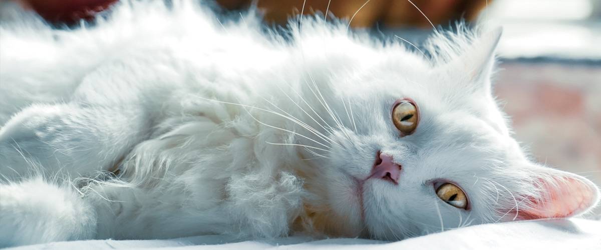
June 8, 2021 – Injection site sarcomas are aggressive tumors located under the skin. They affect roughly 1 in 10,000 cats but can be devastating when they strike. These tumors quickly invade the surrounding tissue and it can be tough for surgeons to know where the tumor ends and normal tissue begins. Dr. Laura Selmic, a veterinary surgeon with a special interest in cancer, talks about a new technology – optical coherence tomography – and how it could be a game changer when it comes to treating these tumors.
0:00:11.5 Dr. Kelly Diehl: Welcome to Fresh Scoop, Episode 33, exploring a new diagnostic tool for use in treating injection-site sarcomas. I'm your host, Dr. Kelly Diehl, Morris Animal Foundation, Senior Director of Science and Communication. And today we'll talk to Dr. Laura Selmic. Dr. Selmic is an Associate Professor in surgical oncology at the Ohio State University College of Veterinary Medicine. So welcome, Laura. Thanks for joining us.
0:00:36.3 Dr. Laura Selmic: Thank you. Yeah. Thanks for inviting me to talk.
0:00:40.3 DD: To start, I always ask everyone, before we dive into your study and a little bit about injection-site sarcomas, can you tell us about your journey to becoming a veterinarian and where you got your veterinary degree?
0:00:55.6 DS: Yeah, yeah. So I grew up in England, and we had a plethora of different animals, anything from dogs and cats to hamsters, rabbits, and later some farm animals. So I grew up, either wanting to be a doctor or a veterinarian, and decided in the end that veterinary medicine was for me. And so I went to the Royal Veterinary College in London to do my vet school, and then I ended up practicing for a couple of years in the UK, and then moving to the US for my residency in surgery.
0:01:32.9 DD: And remind me, where did you do your residency again?
0:01:36.4 DS: I did my residency at Texas A&M, and then did a fellowship at Colorado State University.
0:01:44.2 DD: Which is where I did my residency. And they have a great oncology group there. [chuckle] And it's really strong. I think most people know about it. So you just talked a little bit about this, but why do you think you were drawn to surgery and then specifically also oncologic surgery?
0:02:03.6 DS: Yeah. That's a great question. I, not a hard time in vet school I really enjoyed every single rotation, but my first rotation was actually soft tissue surgery, and it was very inspiring to see how a surgeon could help these animals in need with their skill set. And so that's what attracted me to surgery. One of my initial mentors was Daniel Brockman, who trained at University of Pennsylvania and then is a professor at the Royal Veterinary College currently. So he was a very inspiring, talented soft tissue surgeon. And so during my residency, I really enjoyed the types of surgery we did any soft tissue, orthopedics, neurosurgery, but I was particularly drawn to how the surgery can be part of a bigger team on oncology to be able to really help with integrating surgical treatment of tumors into what is happening with the dog or the cat and what the biology of the tumor is. So it was a very interesting. Claudia Barton was one of the medical oncologists at Texas A&M that was very inspiring.
0:03:18.3 DD: Yeah. It's interesting how we all find our little niches. I think surgery always gave me a lot of anxiety [chuckle] when I was an intern, I was in private practice too, which is probably why I migrated over to internal medicine. So let's move to your project a bit, but I wanted you to kind of get everybody... We have a lot of different people who listen, and I think we have veterinarians and vet students, but we also have some of the lay public. And get us up to speed a little bit on one of the nastiest tumors I think I ever saw in practice, which is injection-site sarcomas in cats. And like when we first started, just the history, because I don't know if we just didn't recognize them for a long time. I certainly saw them when I started in practice, and that seemed to be when in the early '90s, in particular, we were recognizing this. But tell us a little bit more about this really nasty tumor.
0:04:18.3 DS: Yeah, exactly. And so injection-site sarcomas, the kind of history behind it is that it was recognized in the '80s and '90s that cats were getting a very aggressive form of sarcoma, which is a tumor that arises from the connective tissues in the cat, and this was progressing quite aggressively locally. And though initial studies reported a linkage to these developing after vaccinations, specifically vaccinations that contain the compound collagen adjuvant and that was to really increase the potency and response to the vaccine. So subsequent to this, obviously, when they noted this, they've changed the design of vaccines so that it didn't integrate, always integrate an adjuvant there, but actually still these tumors were arising at sites of injection of other compounds as well. So it seems like cats can develop reactions to this sort of minimal kind of injection trauma, which can over a period of months to years develop into this quite aggressive tumor. And this is a little bit in contrast to this tumor, soft tissue sarcoma in dogs, where it doesn't tend to behave as aggressively normally.
0:05:46.7 DD: Just before we get into your study, I want to take a little detour. Is this common... Is this a cat thing? And I don't know the answer to this. Do we see it in other feline species like large cats?
0:06:02.3 DS: Yeah. This hasn't been something that has been reported very extensively in other feline species. It has been most reported in domestic cat species.
0:06:16.5 DD: Okay, thanks. Yeah. That was just a little question that came to my head. So let's start by talking about your study and begin with what you wanted to accomplish with your study, and then we'll get into more detail.
0:06:30.6 DS: So yeah, as a surgeon, we were faced with these aggressive tumors and difficult resections on cats. And so we realized that there was a significant problem that we wanted to try and address, which was we would plan out the surgery based off imaging, like a CT scan or an MRI, and we would remove tissue, but we would have to wait several days before we got a result of the pathology, and then the result would either be we have completely removed the tumor or we have residual potential cells, and after we had finished the surgery, some of these patients would never re-develop a tumor and so would be cured. But some of them, even when we remove the tumor completely would develop tumor recurrence. And so we wanted to see if we could integrate cutting-edge technology to be able to screen cats at the time of surgery for any residual cancer cells to see if we could develop a strategy where we could deal with the cells at the time of surgery, or at least have that as an option. And so we wanted to try and find an accurate method to be able to do this real time. And so this is how the study idea basically came about.
0:07:58.0 DD: And that's a perfect lead-in to talk about your methodology. So if you could start with first of all, how you heard about the technology you used and then a little bit for us lay people on how it works.
0:08:14.6 DS: Yeah. So when I was starting my faculty career after my fellowship, I looked into a lot of different techniques to be able to assess the tissues. One of the techniques that seemed to show a lot of promise was this technique, optical coherence tomography. And so if people have heard of this before, it's normally in the context of scanning of eyes. So it's used very, very commonly for retinal imaging in people. And it basically passes light into the tissue and then looks at the light that's reflected from the tissue to give us an idea about the constituents, the microstructure of the tissues. And so this works very well, and it's become adopted as a gold standard for retinal imaging, and just at the start of my faculty career, they'd been an initial study in human breast cancer that had showed some promise about looking at the surgical margins. So that was what inspired us to look at this technology.
0:09:26.2 DD: So it sounds like this was really early using... You jumped on this, it wasn't really common in people, it sounds like. They were just exploring. Was breast cancer the only thing they were looking at with this?
0:09:39.6 DS: Yeah, exactly. So when we had become interested in optical coherence tomography, this was really only investigational in people and not... There wasn't any FDA-approved device for people nor was there extensive clinical trials in people and different cancer types. So breast cancer was one of the first preliminary clinical studies they had performed.
0:10:09.7 DD: And just really quickly, and I may be dating myself, talk about why some kind of intra-operative examination for this... People have an idea why it would be important, but I remember in my day, sometimes we would do frozens during surgery and you'd zip tissue up to the lab, and I realize not everybody has that in practice. Is that unsatisfying or this is just a easier technology, or why would you look towards this?
0:10:43.0 DS: That's a really great question. So basically, frozen sections are commonly used in human medicine to assess different margin areas. You can only assess a few different margin areas, but it actually also requires a lot of expertise from the pathologist. So that isn't routinely accessible for dogs and cats.
0:11:08.2 DD: So Laura, walk us through the methods, the methodology of your study and how you used OCT in it.
0:11:16.0 DS: Yeah. Great question. So we actually assessed cats that were having feline injection-site sarcomas surgery. So they had their surgery, their surgeon remove the tumor, and then we took the tumor before anything was done with it and scanned it with the OCT. So we looked at every single aspect of the cut edge where the surgeon had cut to look for any footprints of the fact that there might be residual cancer cells to the margins or potentially left behind in the patient. And so because obviously OCT is so new, we compared this actually to what is accepted as the gold standard for assessing surgical margins, which is pathology. So we actually had... After we'd done imaging, without telling the pathologist anything about the results of the imaging, we had... We put the sample in formalin, it fixed, and then the pathologist looked at all of the cut edges as well. So essentially, we did kind of an enhanced pathology assessment of the margins, because normally they only look at a small proportion, but we had them look at the entire margin so that we could compare our results to that.
0:12:31.7 DD: And just for everyone, this instrument is kind of cool. It looks almost like a little ultra... Well, it is a little ultrasound machine. So again, these cats are in surgery and you can just plop this thing sterilely into the surgical site and take a look around. And Laura sent us fabulous pictures which are fun to look at. So moving to the results then, in alluding to the pictures that I've seen, you had a tremendous, great number of publications come out of this study, which is terrific. What did you find starting at the high level?
0:13:10.0 DS: Yeah. So initially we looked at... Within the realms of this study, we looked at actually both cats and dogs with sarcoma to try and look at what the different tissue types at the surgical margins look like on OCT compared to pathology, so that we could look for features of those tissues that we could use to train new observers and the people, the clinicians that will be interpreting these scans in the future, so in the next part of the study. So then in the second part of the study, we took images and videos that we had acquired from imaging all of the cats in the study, and then we had a panel of clinicians of different levels of experience review the images after a training that we gave them to orientate them to what the different tissue types look like with OCT, and then we had them without knowing any of the results of the pathology or anything in-depth about the cases, we have them read those imaging and tell us whether they thought there was cancer present, or whether they thought that the tissues were normal. And so, actually the observers did very well considering they had only a very short training experience of about an hour and a short practice on some images before. They actually had quite a good accuracy for assessing these images. And so we definitely see this as a promising result for a real-time imaging device, which could be utilized hopefully in the future for the real-time assessment in cats with injection-site sarcoma.
0:15:00.0 DD: I think that was one of the things I thought was so cool about your study, was the training aspect and how quickly people could pick this up, because we were talking earlier about pathologists and frozen sections and the training obviously that that requires versus how well folks did with learning how to do this very well in a short period of time.
0:15:27.0 DS: Yeah. I think that we were also very impressed by how quickly the clinicians picked up the differences in tissue types. I think one of the advantages, like you had alluded to earlier is some of the analogies to the way the images look to the way ultrasound looks. And so ultrasound uses sound waves and it's less high resolution than OCT, but it has that same contrast and appearance of the structure of organs, whereas we're looking at a more detailed microscopic level of tissues. But I think some of the interpretation transferred across, which helped the observers in the study.
0:16:08.7 DD: Yeah. It's really mind boggling. So I have a question for you regarding, is there a particular case that sticks out in your mind where this technology really made a difference for a patient?
0:16:26.1 DS: Yeah. I think that's a good question. We did have a couple of really good cases where it was a really large specimen where the pathologist would have had to have been very selective about where they did samples where we could scan and in-depth see some areas that were very concerning for being an incomplete removal or there being residual cancer in that area. And so one of the cases in the study had had some radiation treatment for the procedure, and that it still seemed very clear that we could see the abnormal tissue or the cancerous tissue with the imaging. And so I think that the bigger samples, it really showed us how valuable it is to be able to look at the whole of the margins for this determination.
0:17:19.3 DD: That's awesome. We had a kitty in the study that I know because their owners are donors, Oyl, O-Y-L.
0:17:26.8 DS: Yeah, Oyl. Yeah.
0:17:29.4 DD: Yeah. And they... I think that kitty went on other than developing grey hair, I think where it used to be black a kitty it had a patch of grey, did marvelously well with...
0:17:41.8 DS: Yeah. Oyl was one of the first cases that we involved in this study, and yeah, I have some great pictures initially post-op of Oyl in a little jacket to try and cover the site where the cat had surgery. So yeah. Sweet cat.
0:18:01.9 DD: Yeah, really great. That was a great outcome, I think. So I have a question maybe that you... Would be tough for you to answer. As this technology, like all technology, advances, is it... Do you see it becoming cost-effective for beyond just a large university, for example a referral practice like what I worked in or a big private practice?
0:18:27.4 DS: Yeah. That's it. It's a really good question, and I think... Certainly, I think that this is probably quite analogous to the way that ultrasound came into clinical use and veterinary medicine where at first the systems are kind of bigger and they're more expensive, and then as more companies develop and utilize this technology it becomes an economy of scale and so it becomes more cost-effective. The system at the moment, the research system that we are using was about $60,000. So it's not extremely high and out of the realm of VM to use, but I imagine that when companies are producing these mainstream that that cost could be significantly lower as well.
0:19:17.4 DD: That would be awesome. When you said that, I can remember, which now I will date myself, our first ultrasound machine, which was like $30,000 bucks, it was this huge machine with a little tiny screen. And then by the time I retired, it was an easy, little wheel around unit with a giant screen and all these capabilities. So I'm excited that this will get better. So just for you, what are projects that you're working on now. It doesn't have to be with OCT, but are you looking at more things with OCT and where next? What are you doing next?
0:19:52.5 DS: Yeah, thanks. That's a great question. So at the moment, we have just finished a project looking at OCT for any skin or subcutaneous tumor in dogs, and that was a fun project to be able to complete looking at a more kind of heterogeneous population of patients with different types of masses. And we are just collating the results of that study, but it seems encouraging as well in that patient population that OCT was able to be interpreted with a high accuracy, again, which is good. We're working at the moment, we started actually a human clinical trial with physicians at OSU Wexner Medical Center, the James Cancer Hospital, where we are actually starting to look at a preliminary study with human soft tissue sarcoma patients, looking at their surgical margins after their tumors are removed just like we did with cats.
0:20:51.7 DS: And so the information that we found out in the cat study has directly informed as looking at this with human patients. So that's exciting. We're hoping to apply for some grant funding to make it into a bigger clinical trial in people, which would be really cool. And then we are actually looking at starting some projects with a new type of OCT, which is a set of the hardware and software modifications, so OCT to look at the polarization of the light in addition to the light that's back-scattered by the tissues. And so we're hopeful that that will help us even further increase the accuracy with giving us more information about the collagen structure within the tissues. We see that as a promising area to look into.
0:21:46.5 DD: That sounds good. It sounds like it could be used maybe for other things, because you're looking at just really tissue... I'm thinking of even like horse tendons, which was the other thing that really some fine resolution helps with that, but it sounds really awesome. So if I can pin you down with, what would your take home message be for veterinarians who are out there, vet techs and owners who are faced with injection-site sarcoma in cats?
0:22:14.1 DS: Yeah. That's a really good question. I guess I would say that it's good for us to take these tumors very seriously from a perspective that when they are detected early diagnosis and then well-planned treatment using advanced imaging and then a more aggressive surgical resection seems to be in the best interest of these patients often combining with radiation therapy, we can get really good outcomes for these patients would definitely deter doing closer excisions or just removing it to find out what it is with this aggressive disease.
0:22:55.9 DD: So it sounds like, again, don't take any lumps or bumps on a cat, even if it's not vaccine, I think you've made a good point, and it's not necessarily vaccinations, it can be any injection, and it can be the 100th time they get the same injection, that we should be paying attention to those. So that's really great advice, I think, for everyone. So that does it for us. For another episode of Fresh Scoop. So once again, thanks to Dr. Laura Selmic for joining us.
0:23:29.1 DS: Thank you very much, I appreciate the opportunity to chat.
0:23:33.0 DD: So everyone, we'll be back with another episode next month that we hope you'll find just as informative. And as we all know, the science of animal health is ever-changing and veterinarians need cutting-edge research information to give their patients the best possible care, and of course, that's why we're here. You can find us on iTunes, Spotify, Google Podcasts and Stitcher, and if you like today's episode, we'd sure appreciate it if you could take a moment to rate us because of course, that will help others find our podcast. And as always to learn more about Morris Animal Foundation's work, go to morrisanimalfoundation.org, and there you'll see just how we bridge science and resources to advance the health of animals. And you can also follow us on Facebook, Twitter, and Instagram. And I'm Dr. Kelly Diehl and we'll talk soon.


