DENVER/June 29, 2021 – Morris Animal Foundation is funding researchers at the University of Central Lancashire, alongside colleagues at Utrecht University and Delsys Inc., to quantify how a horse’s muscles and limb movements adjust to accommodate lameness. The findings of this study will provide a greater understanding of the clinical signs of lameness, which could guide future diagnostics and treatment.
Lameness is a change in gait, often to relieve pain in a limb or the back, and is the most common reason for veterinary examination. Current diagnostic methods for lameness rely heavily on subjective assessment, including observing how the horse is weight shifting. The mechanism of shifting weight from one limb to another is assumed to require muscle contraction and coordination adaptations. Still, these neuromuscular changes have yet to be measured or described.
“We know that horses alter their movement pattern when they’re lame, but we don’t know much about the functional changes in muscles that facilitate these changes in movement,” said Dr. Lindsay St. George, Research Fellow at UCLan, and the study’s primary investigator. “We want to define muscle activity in clinically sound, non-lame horses, and then use this knowledge to quantify adaptive changes in muscle activity that occur when a horse is lame.”
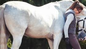
St. George’s team uses surface electromyography (sEMG) to quantify muscle function and 3D motion capture technology to quantify movement in horses. sEMG is a non-invasive technology that measures muscle activation by recording the electrical activity produced by skeletal muscles when they contract. This study employed Delsys Trigno sEMG sensors, which are small, wireless sensors attached to the horse’s skin over-lying superficial muscles of interest.
St. George and her Utrecht colleagues previously evaluated eight horses at that university. They placed sEMG electrodes over selected muscles and reflective kinematic markers on each horse’s forelimbs, hindlimbs and back. Each horse was then trotted down a hard surface runway. The data collected from these trials were used to establish a baseline for each horse’s movement and muscle activity patterns when they were clinically sound.
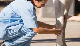
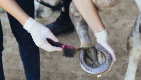
Then, veterinarians induced mild, temporary lameness by applying pressure to the sole, using a modified horseshoe technique. This commonly is employed in research to standardize lameness. Left or right forelimb lameness was randomly induced, and horses were trotted to collect data. After a minimum of 24 hours and ensuring the horses did not show residual lameness, sEMG and kinematic data were collected again from baseline and hindlimb lameness conditions.
Now the team is analyzing the data, looking for differences in the activation timings and amplitude of the sEMG and kinematic signals between the conditions. Analyzing both sEMG and 3D kinematic data will allow the researchers to accurately measure the relationships between changes in muscle function and movement during lameness.
“Lameness is one of the most common problems we see in horses, but we still have a lot to learn about diagnosis and treatment,” said Dr. Janet Patterson-Kane, Morris Animal Foundation Chief Scientific Officer. “If this technique will help us objectively measure the true condition of all of an animal’s musculoskeletal tissues, it will assist in optimizing treatment on an individual basis.”
If her study is successful, St. George would like to conduct a similar survey, but collect data from a larger group of horses on different muscle groups and clinical lameness cases.
About Morris Animal Foundation
Morris Animal Foundation’s mission is to bridge science and resources to advance the health of animals. Headquartered in Denver, and founded in 1948, it is one of the largest nonprofit animal health research organizations in the world, funding more than $136 million in critical studies across a broad range of species. Learn more at morrisanimalfoundation.org.
###
